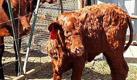
Cattle owners sometimes encounter unusual symptoms in cows, namely, cones on their body, which can occur in a variety of places: on the udder and abdomen, back and neck, jaw and other areas. This sign must be alerted, because most often the bumps on the body of the animal are manifestations of serious diseases.
General description of the problem
Subcutaneous bulges themselves are not an independent disease — it is only a symptom by which a disease can be diagnosed. Thus, there are several serious pathologies of allergic or infectious origin. For an accurate diagnosis, a qualified veterinarian is required.
Cones can occur in cows in different parts of the body, and their localization is partly indicative of exactly what hurt the animal. They may vary in size and shape, as well as in color, density, and palpable consistency. The number of formations can also be different and vary from single manifestations with a clear localization to a massive lesion of the whole body with diffuse propagation.
Did you know? Sunshine is vital to cows, because their bodies cannot break down vitamin D without it.
Most often, growths on the body are not the only symptom and may be accompanied by a number of other manifestations - fever, lethargy, lack of appetite, and others. However, this is the brightest and most noticeable signal that not everything is in order with the animals.
Of course, there are cases when the formations are relatively harmless - for example, mechanical damage to the skin or a sucked tick. However, even in these cases, you should not neglect the concern for the health of the pet, because the mechanical damage can fester, and the tick bite can lead to some blood loss.  In addition, ticks are carriers of many dangerous and deadly diseases. Therefore, when detecting dubious growths of any shape, size and location, it is best to immediately isolate the cow from the rest of the herd, call a specialist, and examine and treat only with gloves and a mask.
We recommend reading about how to put injections to cows and calves.
What may be hiding under the bump
The causes of such formations are infectious, parasitic and viral diseases, and sometimes an allergic reaction. All of these conditions require immediate treatment.
Allergy
Allergy - This is an increased pathological immune reaction of the body to an allergen, which is a harmless substance, but which the animal's organism mistakenly recognizes as dangerous and reacts according to the type of protective mechanism.  Causes:
- Feed. It can be not only non-certified and low-quality feed from the manufacturer, but also food of natural origin - a certain grass and everything that a cow could eat on free grazing. However, one-time use rarely has such serious manifestations. Usually, in order for a life-threatening allergic reaction to occur, contact with the allergen must be constant and regular. Therefore, it is necessary to revise the diet of the animal and eliminate all questionable foods.
- Supplements. Substandard supplements that do not undergo the necessary cleansing and certification can cause an allergic reaction.
- Medication. Drugs and vaccines are designed to cure one disease, but can cause another - allergies, especially if the medication was administered with a violation of the instructions or without the appointment of a veterinarian.
- Repellents and insecticides. Not all such drugs are safe, and they can cause allergies, as their composition is quite volatile and aggressive.
- Means for cleaning, which is processed barn. The chemical composition of such agents is a strong allergen, especially if the remnants of detergent preparations were not thoroughly washed out and the animal is in direct contact with a hazardous substance.
- Any other substances in the environment. It can be whitewashed in the barn, bedding, wood walls and floors, the materials of which are made of feeders, drinkers, buckets and any other inventory for the care of livestock.
Read more about the causes, symptoms and methods of treatment of allergies in cattle.
Places of manifestation
The spread of cones on the body of a cow may not have a clear localization. The formations are small, most often small, the size of a pea, spread throughout the body under the skin. First, the growths are rare, but if the contact with the allergen does not stop, they quickly spread and can cover all parts of the body.  Related symptoms - tearing, profuse mucus in the nose, sneezing, coughing, urticaria, red eyes and throat, plaque on the tongue. Also, allergies can be accompanied by the spread of large spots on the skin, the fur becomes erect, edemas of various localization and volume occur, breathing becomes heavy and frequent, and the heartbeat becomes more frequent.
Important! Allergic reactions are most dangerous for the occurrence of anaphylactic shock, which is likely to be fatal. Therefore, when a significant body edema is found in a cow and breathing is difficult, it is necessary to respond promptly.
Treatment
First of all, antihistamine (antiallergic) drugs are prescribed to the animal: Dimedrol, Diprazin, Hydrocortisone or others. The route of administration is intramuscular. You also need to urgently identify the source of the allergen and isolate the cow from it. If the reason is in the feed, then the animal is transferred to a strict diet.
If the source lies in the environment, then the cow is transferred to a new room. But if the cause of the allergy is not clear, then take all precautions - diet, the absence of any chemicals, clean and hypoallergenic content. Additionally, the veterinarian may prescribe a course of vitamins to maintain the immunity of the cow, which contributes to the development of an adequate body response to the allergen. 
Actinomycosis
Infectious fungal disease, the source of which is the defeat of an animal with radiant fungi. Infection is possible throughout the year, as the fungus is very resistant to temperatures and environmental conditions.
Causes
Radiant fungi often enter the body of livestock through contaminated feed, hay and water, and can be transmitted from other sick animals. However, a prerequisite for their penetration into the body is the presence of damage on the mucous membranes or the epithelial layer of the skin.
Did you know? Each cubic centimeter of a cow's stomach contains over a million single-celled organisms. - ciliates. With the help of such “cohabitants”, cattle are able to digest the rigid cell walls of plant foods.
That is why cows are sick with actinomycosis most often in the winter period, because just at this time they consume roughage that damages and scratches the oral mucosa.
Actinomycosis often affects calves during teething. However, adults are no less susceptible to this disease. The fungus gets through any damage - scratches on the mucous membranes of the mouth and nasopharynx, wounds in the gastrointestinal tract, cracked nipples, injured on the surface of the skin.  Places of manifestation
The first symptom of the disease is the formation of large bumps. Localization depends largely on the place of penetration of the fungus in the tissue. Since damage to the mucous membranes is most likely, this causes the greatest frequency of occurrence of cones in the area of the head and jaw.
From the tissues of the mucous membranes, fungi migrate to the lymph nodes in the neck, where they actively proliferate and therefore cause the formation of growths in the neck. But bumps can also occur on other parts of the body where the fungus could get.
Cones are single, local, in size can be very large. Palpation feels slightly colder than the skin in nearby areas of the body. Dense in consistency. Galls quickly increase in size, grow together with the nearest healthy integuments. If localization is on the head, then the head shape of the cattle will gradually change, distort.
Over time, actinomycomas (bumps) are increasingly filled with exudate and purulent contents, soften, grow, become mobile and soft. After aging, they erupt in the form of fistulas - holes appear, through which purulent contents ooze out.
For a long time, the disease can occur in a chronic form, and its incubation period lasts from several weeks to a year. At this time there are no clinical signs.  Related symptoms
The temperature of sick animals usually does not increase, and its increase may be triggered by the acceding secondary infection. But actinomycoms make life difficult for cows. For example, when formations occur in the jaw, pharynx, and neck, they make chewing and breathing more difficult, which causes cattle to quickly lose weight.
If the bones of the skull are affected by actinomycosis, then the structure of the head changes, as the affected areas increase and become twice as thick. Also often an additional symptom is tooth loss. With the defeat of the udder, the fistulas practically do not heal due to the fact that the tissues of this organ are very sensitive. Tumors are localized mainly in the posterior lobes of the udder and are accompanied by skin necrosis.
In rare cases, the causative agent of actinomycosis can migrate to internal organs, affecting the gastrointestinal tract, bones, kidneys and liver. This happens with a low immune resistance of the body, and in this case the chance of a successful cure is minimal.
Learn more about the treatment of actinomycosis in cows.
Treatment
The diseased individual is isolated from the rest of the herd to avoid further spread of the infection. It is transferred to a clean room, the processing of which can be done with a 3% solution of freshly sour lime. Further treatment depends on the stage of the disease.  In the early stages, it is enough to give the sick cow an injection of potassium or sodium iodide. Injections are made both intravenously in the form of prokapyvany, and directly into the affected area. If these measures are taken at the very beginning of the disease, then additional therapy may not be necessary. If the treatment is started late, then antibiotic therapy is necessarily attached to iodine preparations.
A veterinarian may prescribe a course of antibiotics - amoxicillin, oxytetracycline, polymyxin, benzylpenicillin. To the last drug resort only in the most severe cases. With low effectiveness of antibiotic treatment, the animal may be offered surgery: growths are opened, and the affected tissues are removed.
Important! Antibiotics are continued even after all symptoms have disappeared. This is necessary so that the infection does not have a relapse.
Bursitis
This disease is an inflammation of the synovial bags (bursa), which are a small closed cavity around the joints and are formed by connective tissue, the fibers of which, due to mechanical damage, stretch and move. Bursa inflamed, filled with exudate, which leads to the formation of large cones with watery content.
Causes
Inflammation of the synovial bags around the joints most often occurs due to any closed and open injuries of these parts of the limbs. Such an injury can cause joint injury. 
Risk factors are the following circumstances:
- cows are kept crowded;
- too short leash of an animal without the possibility of movement;
- hard (stone, reinforced concrete) cold floors without bedding, insulation and softening.
Familiarize yourself with the symptoms and treatment of bursitis of cattle.
Usually the bursa contains pathogenic microorganisms, but for a long time they are in a latent state. Bruising and tissue damage activates their vital activity, which is why inflammation occurs.
Less commonly, bursitis develops due to infection and decay of adjacent tissues, when the infection penetrates into the synovial sac, causing inflammation. In very rare cases, the disease may occur as a secondary consequence of diseases such as sepsis, tuberculosis, brucellosis, and others.
However, not every bruise or primary infection leads to bursitis. The onset of the pathological inflammatory process in synovial bags is often preceded by a general depletion of the animal, low body resistance to environmental factors and resistance of tissues to mechanical damage.
Places of manifestation - predominantly limbs of cattle. Leg joints are affected - most often carpal or knee.  Inflammation of other joints of the body is possible, but this is extremely rare, because the legs of animals are most susceptible to mechanical damage. On the legs around the joint with an inflamed bursa a large mobile bump appears, filled with fluid.
Related symptoms
Because of the painfulness of the injured limb, a cow has lameness - from mild to severe and very noticeable, sometimes the animal cannot even move due to pain. The resulting tumor prevents flexion of the joint, which significantly interferes with movement. Sore limb may shake and shake. The swelling to the touch is hotter than the rest of the body.
The acute form of purulent bursitis may be accompanied by fever, weakness, lethargy, severe pain. However, the appearance of purulent content does not occur immediately. At the beginning of the disease, the synovial bag is filled with serous contents, then with serous-fibrous, and, if no treatment is provided by this point, bursitis becomes purulent.
The disease has a chronic and acute form. In acute form, the lump is painful and soft, filled with exudate. In the chronic form, keratinization of the skin and subcutaneous tissues occurs, the synovial sac fuses with the joint, making it immobile and causing severe lameness.
Did you know? Cows go to rest quite often: during the day they can go to bed and rise up to 14 times, and sleep in fits and starts for 1.5-2 minutes.
Also, depending on the type of content of the bursa, there are aseptic and purulent bursitis. If the disease becomes chronic, then a fistula appears on the surface of the lump, from which pus constantly oozes.
Treatment
First of all, it is necessary to eliminate the cause that led to the occurrence of bursitis. The cow is given a rest, placed in a warm room on a soft bed. Treatment can be both conservative and operative.
The latter is usually resorted to with keratinization of the bursa and its accretion with the bones and the joint, as well as with the accumulation of an excessive amount of pus with the subsequent formation of fistulas. If the disease is a secondary manifestation of the underlying infection, then treatment measures should be primarily aimed at its elimination. In other cases, treatment is usually local.
Video: bursitis treatment in a cow At the initial stage, compresses are applied to the swelling, designed to relieve inflammation and tenderness, for example, alcohol, or make antiseptic dressings with an ointment containing an antibiotic. The affected place can be irradiated with a blue lamp, apply hot or cold heaters.
Antibiotics are often injected into the injured area. It may be such drugs as "Ceftriaxone", "Amoxil", "Gentamicin" and others. The skin is treated with hydrocortisone ointment, designed to relieve inflammation. If the disease has passed into that stage when too much pus is formed, or the lump has burst in the form of a fistula, then surgical intervention is required.
The inflamed bursa is anesthetized with novocaine, pierced, freed from pus, scraped, disinfection is performed with "Furacilin" or "Sulfacil", and then filled with a tampon soaked with cauterizing solution. An antibiotic is injected around the affected area.
Самолечение бурсита недопустимо: некорректное лечение приводит к переходу заболевания в хроническую форму с последующим ороговением синовиальной сумки и участков вокруг неё, когда исправить последствия возможно только хирургическим удалением ороговевших тканей.
Гиподерматоз
Это заболевание относится к паразитарным, его провоцирует подкожный овод. It has a chronic course and is very dangerous for cattle, as it makes meat and milk unsuitable for consumption.
Learn more about cattle hypodermatosis.
In places of localization of gadfly larvae, dry, clearly palpable bumps appear. 
Causes
Hypodermatosis provokes a hypodermic gadfly, namely, its two types that are common in the Northern Hemisphere are the ordinary subcutaneous and southern subcutaneous gadflies.
Important! The fact that cows are affected by parasites can be suspected by the strange "cooperation" of cattle with wild birds. Sparrows, jackdaws and magpies sit on the back of animals and peck out from under the skin of gadflies. In half an hour, birds can peck up to 400 gadflies.
Insects infect cows predominantly in summer during flight. The larvae migrate to the esophagus and the spinal canal, infect the internal organs, often localized under the skin in the form of hard and dry small bumps.
After the larvae have settled under the skin, capsules appear around them, which is clearly and palpably defined as a bump under the skin of a cow. Over time, a hole appears on the bump from which exudate oozes. It is through this fistula that the gadfly larva will leave its owner in order to form a pupa on earth and in a month and a half turn into an adult gadfly.
Young cows and bull-calves are most susceptible to hypodermatosis, since their skin is quite tender and is not an obstacle to the penetration of the larvae, and the immune response is too weak to fight the parasites.  The emergence of cones - the most obvious stage of the disease. Subcutaneous formations with palpation are felt hot, for an animal they are very painful.
Important! The first six months after infection, the disease has a hidden form, so there is a high risk of buying sick individuals or the spread of the parasite among the herd due to unidentified sick individuals.
Places of manifestation
Most often, bumps with capsulated larvae appear on the back, lower back and sides. However, most often the main place of manifestation is the back and its areas adjacent to the spine. Cones can also occur on the sacrum, chest and neck, very rarely occur on the lower abdomen and udder or on the head. Observed small encapsulated seals in the esophagus and spinal canal.
Related symptoms
Ill cattle become very restless, nervous, tossing and rubbing, itching. The migration routes of the larvae are also very painful for cows when pressed. There is a general depletion of animals, there are signs of a general intoxication of the organism, provoked by toxic activity of the gadfly larvae.  When they die, even more toxins enter the bloodstream, poisoning the sick individual. Necrotic decomposition of the skin and subcutaneous tissues may begin in the surrounding areas. The general depletion of the body from intoxication leads to a decrease in the productivity of cattle.
Important! If the larvae are too large in the spinal canal, paralysis of the hind limbs may occur, and if there is an accumulation in the esophagus, problems with chewing and swallowing occur.
Treatment
consists of chemotherapy with insecticides. Moreover, they distinguish early chemotherapy, which is carried out at the first stage of infection by the larvae - around the beginning of September, immediately after the end of the insect flight period.
But these measures are more likely to be preventive in nature, since it is almost impossible to make an accurate diagnosis and identify sick individuals because of the implicit clinical picture. And the second type of chemotherapy is late. It is carried out at the final stage of development of the larvae, when they are already localized under the skin in the form of cones. Carcasses of non-surviving cows are disposed of to prevent the spread of the epidemic.
Nodular dermatitis
This disease is also widely known as a cusp. It is a dangerous viral disease, in which every tenth animal dies without treatment, and with appropriate treatment, the lethal outcome is reduced to 1-2%.  Patients individuals on the body appear numerous cones of different sizes - from 0.5 mm to 7 cm in diameter, they are flat and their height does not exceed 0.5 mm. The number of formations can be so great that they merge into formless subcutaneous inflammation.
Also a distinctive feature is flaking of the skin along the perimeter of the cones, the formation of a fistula in the middle of the growth, in the center of which there are incorrectly growing hairs of different color. 7–20 days after the appearance of the first formations, an easily detachable tube of necrotized tissue appears in their center, which may itself disappear.
The incubation period can last 3-30 days, although most often it does not exceed a week. The body’s immune response to virus penetration occurs 2-5 days after infection, when the first bumps appear on the skin.
Causes
Until recent years, the disease was considered exotic, African, but now it is increasingly common in our countries. It is spread mainly by blood-sucking insects, although infection occurs through direct contact with sick animals, through household items and feed. The virus strain is very resistant, suffers a threefold freezing and is not completely destroyed during antiseptic treatment of the premises. 
Places of manifestation
There is no clear localization of cones. They are numerous, of different sizes, occur throughout the body in different quantities, which depends on the extent of the disease. In sick calves, the bumps may not be observed at all, and the only manifestations of the tubercle are diarrhea and fever.
The defeat of the subcutaneous layer is not the only manifestation of nodular dermatitis. The disease affects the mucous membranes of the nasopharynx, genital organs, lymph nodes, the gastrointestinal tract and the respiratory system.
Related symptoms:
- fever, accompanied by an increase in body temperature to 40-41 ° C;
- lymph nodes are significantly increased in size, painful and sedentary;
- from the nasal passages oozing mucus with an unpleasant odor;
- eyes too watery;
- increased salivation;
- when udder is damaged, the milk becomes pink, it is difficult to stand out, and when heated it turns into a gel;
- lethargy and weakness;
- refusal of food, lack of appetite;
- decrease in productivity in the form of reduced milk production, possible agalactia, loss of body weight.

When the disease is neglected, respiratory organs and the digestive tract are affected, and ulcers are found not only on the skin, but also on the mucous membranes of the mouth, nasopharynx, and vagina in cows.
Treatment
Nodular dermatitis spreads massively and affects about 90% of the entire herd. Patients are quarantined and kept in it for a month after the disappearance of the last clinical signs of the disease. Bulls are kept in quarantine for at least 2 months, since during this time the virus remains active in their sperm.
We advise you to read about how to treat nodular dermatitis in cattle.
Special methods of prevention and treatment of the tubercula have not yet been identified. Recovery mainly occurs independently, and therapeutic measures are manifested by symptomatic therapy aimed at preventing severe complications such as pneumonia in the adult herd and enteritis in the calves.
Treatment can be carried out with the introduction of antiviral drugs "Bifer-B", "Gentabiferon-B" and "Enrofloksavetferon-B". To prevent severe cases of enteritis in young animals, strong antibiotics (for example, tetracycline) are used.
Video: nodular dermatitis in cattle The cattle requires careful care, it must be provided with high-quality food and good conditions of detention. Sick livestock is regularly bathed in a disinfectant shower, treated with aerosol preparations, which is especially effective for preventing pulmonary complications. Ulcers are treated with bactericidal ointments - Vishnevsky, zinc, syntomycin.
Smallpox
This dangerous virus is quite rare lately, as mankind has found effective ways to combat it. Ospovirus affects not only cows, but other animals as well as humans.
Causes
In two cases, livestock may get sick with smallpox — when a vaccinia virus is transmitted from newly vaccinated personnel or with a true smallpox virus that is transmitted by insects, rats and mice, and other sick animals.
Did you know? Dr. Edward Jennette invented the first smallpox vaccine for humans: it was created on the basis of cowpox. In memory of the doctor's vaccinations were called vaccines from the Latin "vacca" - "cow".
Smallpox is most often affected by young individuals with an unformed immunity or adult livestock suffering from hypovitaminosis. The virus spreads very quickly and can affect the whole herd and people who are in contact with it.  Infection occurs predominantly through contact with saliva and mucus from the nose of a sick animal, as well as through direct tactile contact with the affected skin areas and falling away dry crusts from the lesions. Ovspovirus penetrates the body most often through the mucous membranes and nipples, abrasions and scratches on the body.
However, with vitamin A hypovitaminosis, when the body's defenses are weakened, the virus can penetrate through whole skin. The incubation period lasts from 3 to 9 days. In those places where the virus has penetrated into the cells, inflammatory processes begin.
First there are focal reddening - roseola, which after a couple of days become bubbles with liquid contents - papules, and even after a while they transform into pustules. From the affected skin, the virus migrates to the internal organs, into the blood and lymph nodes. The latter significantly increase in volume and redden.
Animals with good immunity suffer from smallpox in a mild form - not many papules arise, they are limited to partial necrosis, they dry out and form crusts. After the scabs fall off, the skin itself is restored.
If the course of the disease is uncomplicated, then recovery occurs after 20-28 days after the onset of the first clinical symptoms, and with the appearance of complications it is delayed up to 2 months. If cows are affected by vaccinia virus, the course of the disease is easier and shorter, since only superficial skin is affected. 
Places of manifestation
In cows, papules appear mainly on the udder, milking and stomach. In bulls - on the stomach and scrotum. In rare cases, it may occur in other parts of the body. In calves, the mucous membranes of the nose and pharynx are often affected. The number of "bumps" can vary from single to a couple dozen, depending on the severity of the disease.
Related symptoms
Before the appearance of papules, lethargy of the animal, loss of appetite or its loss, and a decrease in the amount of milk yield indicate the beginning of the disease. Soon the cattle comes a short-term increase in temperature to 40-41 ° C, after which the first bubbles appear.
With the defeat of the udder, it becomes dense, painful. The cow tries to arrange the hind limbs as wide as possible so as not to touch the udder with them - because of this, its movement is impeded. Milking is painful for an animal, milk is poorly excreted, lactation in some cases may stop altogether.
The diseased individual becomes restless, sometimes aggressive, does not allow the milkmaids to come to him. Udder looks puffy and reddened. In young animals, the disease is often accompanied by gastroenteritis and bronchopneumonia. 
If the animal’s immunity is very weak, then secondary bacterial infections join the smallpox. In this case, the prognosis for recovery is dubious. With severe complications, the smallpox virus infects many internal organs, causes disintegration and cell changes, and organ rotting.
Thus, the heart muscles become flabby, the spleen grows, putrefactive processes begin in the liver, the respiratory system and lymph nodes are affected, and ulcerous rashes can be observed not only on the skin, but also on all mucous membranes, in the internal systems and organs. In this case, the animal most often dies.
Treatment
Self-treatment of smallpox is unacceptable, as it may be more harmful to the animal. First of all, sick individuals must be isolated from the rest of the herd to prevent the spread of the virus. Despite the fact that cows in this state are poorly eaten, they still need to be provided with enough food, it is possible to give semi-liquid light food.
It will be helpful for you to read about how to treat smallpox in a cow for an udder.
The room for maintenance should be warm and dry, with good ventilation. Cows must be sown regularly to prevent the development of mastitis. In extreme cases, if milking is not possible, a catheter is placed in the udder of the cows in order to facilitate the outflow of milk.  A definite treatment regimen for smallpox in cows still does not exist. Basically it is symptomatic. Antibiotic therapy is often carried out, since secondary bacterial infections begin to develop in a weakened body.
Important! Milk of smallpox-infected cows is unsuitable for consumption. It is disinfected, and then disposed of as waste.
Local treatment of papules and pustules is also carried out. They are cauterized and dried with Burov's liquid, tincture of iodine, boric acid solution or 3% chloramine. Crusts soften fat and glycerin ointments. For faster healing, the damaged areas are treated with boric, salicylic, zinc or petroleum jelly.
Irrigation and washing of the nose with a solution of boric acid. As we can see, the bumps appearing on the body of a cow are a symptom of serious diseases. They differ in type, size, number and location. Their treatment depends on the cause of the disease, and it can only be carried out under the supervision of the veterinarian who established it.